![Medical News Asia | Powered by ['mediPr]](https://medi-paper.com/wp-content/uploads/2020/02/MediPR-MediPaper-Medical-Communications-Ltd-Healthcare-Writers-Medical-Writers-Medical-Writer-Agency-Hong-Kong-Medical-Writing-Services-Solutions-Quote-醫學寫作-香港-醫學作家香港-medical-PR-public-relations-1030x374.png)

OPHTHALMOLOGY
HK Ocular Surface Newsletter Issue No. 2 Mar 2023
Last updated: 27 March 2023 | In: Ophthalmology, Therapeutic Areas
Article Keywords
dry eye disease, meibomian gland dysfunction, ophthalmology, topical cyclosporine, vernal keratoconjunctivitis

DR. ARTHUR C.K. CHENG
Specialist in Ophthalmology
Hong Kong Sanatorium & Hospital
The Right Timing to Start Cyclosporine in Allergic Conjunctivitis
Atopic keratoconjunctivitis (AKC) is a chronic ocular inflammation commonly diagnosed in patients with atopic dermatiatis. The main symptom of AKC is bilateral itching with frequent blinking, which is mostly observed in children. Other clinical manifestations include papillae on the conjunctiva and eczematoid changes on the periorbital skin. Sight-threatening complications, including corneal ulceration and bacterial keratitis, may occur as the disease progresses.1-31. Bielory B & Bielory L. Atopic dermatitis and keratoconjunctivitis. Immunol Allergy Clin North Am. 2010;30(3):323-36.2. Yang B, et al. Quantitative evaluation of lipid layer thickness and blinking in children with allergic conjunctivitis. Graefes Arch Clin Exp Ophthalmol. 2021;259(9):2795-805.3. Power WJ, et al. Long-term follow-up of patients with atopic keratoconjunctivitis. Ophthalmology. 1998;105(4):637-42.
Vernal keratoconjunctivitis (VKC) is another ocular inflammatory disease considered to be highly similar to AKC. Both AKC and VKC can occur in children and young adolescents. Although it is generally acknowledged that AKC tends to be perennial and VKC tends to be seasonal, VKC-related inflammation with recurrences throughout the year was reported in about one-quarter of patients.4,54. Kim SE, et al. Systemic interventions for severe atopic and vernal keratoconjunctivitis in children and young people up to the age of 16 years. Cochrane Database Syst Rev. 2020;10(10):CD013298.5. Ukponmwan CU. Vernal keratoconjunctivitis in Nigerians: 109 consecutive cases. Trop Doct. 2003;33(4):242-5. “Sometimes, it could be challenging to distinguish AKC from VKC by diagnosis because the two conditions share very similar clinical manifestations. The classification based on age and seasonal variations may not be practical,” said Dr. Cheng.
Dr. Cheng shared his routine practice, starting with the categorization of patients with allergic conjunctivitis (AC) based on the presence or absence of symptoms and signs. Patients with symptoms only usually have less inflammation and often experience ocular redness and itchiness, as well as increased frequency of eye rubbing and forced blinking. Patients presenting with symptoms and signs may have the following clinical features: inflammation of conjunctiva, limbal Horner-Trantas dots, follicles inside the inferior eyelid, and corneal involvement. These two groupings serve the purpose of treating patients with different disease severities differently.
Dr. Cheng’s treatment goals of his symptom-only AC patients are symptom relief, reduction of exacerbations and maintaining remission.66. Hossain IT, et al. Pharmacotherapeutic management of atopic keratoconjunctivitis. Expert Opin Pharmacother. 2020;21(14):1761-9. Management will include avoiding exposure to allergens, and using artificial tears and antihistamine medication for symptom relief and minimizing inflammation. For patients with AC symptoms and signs, the treatment goal is to prevent complications induced by inflammation. Dr. Cheng mentioned artificial tears and antihistamines are generally ineffective to suppress inflammation in patients with persistent signs. Therefore, topical corticosteroids and topical cyclosporine (CsA) could be considered in these cases.
“When patients show prominent inflammation and clear allergic signs, for example, papillae and limbitis, both topical CsA and topical corticosteroid should be prescribed concomitantly QID for a week. After a week, the steroid could be completely taken over by topical CsA, and CsA monotherapy could be continued for the rest of the treatment course,” Dr. Cheng highlighted. Dr. Cheng added that QID topical corticosteroids like prednisolone acetate 1% are fast-acting within 2 days, while topical CsA takes a few days to a week to be effective. Also, topical CsA is relatively safe for long-term use. “By the time of prescribing corticosteroids is needed, topical CsA should be prescribed for my patients at the same time. These patients, no matter diagnosed with AKC or VKC, seldom have their symptoms completely resolved after 1-week corticosteroid treatment. Thus, the use of CsA has a steroid-sparing effect,” Dr. Cheng explained.
Case Sharing
A 12-year-old boy with frequent eye blinking who was pre-treated with oral and topical antihistamines was referred to Dr. Cheng. The patient had no known medical history. Prior intervention with antihistamines failed to resolve his symptoms. Slit-lamp examination revealed limbitis and conjunctival papillae in the affected eye (Figure 1). Mild staining was presented. The case required immediate treatment due to prominent signs of inflammation.
The patient was prescribed with topical corticosteroid (Q2H on the first day and QID on the second day) and topical 0.1% CsA QID right away. During the follow-up visit, significant improvement was seen after a 1-week treatment (Figure 2). The redness and conjunctival papillae had faded completely. Topical corticosteroid therapy was then stopped and the patient was continued on CsA treatment. Dr. Cheng tapered down topical 0.1% CsA from QID to BID, and eventually to QD as the condition improved.

Figure 1. Affected eye before treatment
Figure 2. Affected eye after one-week combination therapy of topical 0.1% CsA and topical corticosteroid
Key Takeaways
It is important to recognize AKC and VKC are not only allergic diseases, but also inflammatory diseases at a later stage. The key to AC management is the timely control of inflammation. Artificial tears and antihistamines are not effective enough in controlling inflammation. The prescription of topical CsA bridging with topical corticosteroids concomitantly should be considered in patients presenting symptoms and signs of inflammation, or in patients failing artificial tears and antihistamines.

DR. JACKY W.Y. LEE
Specialist in Ophthalmology
C-MER Eye Center
Anti-glaucoma medication-induced Ocular Surface Disease: From Cause to Management
Glaucoma is a chronic, progressive optic neuropathy where intraocular pressure (IOP) lowering remains the main modifiable factor for slowing down disease progression. Thus, patients often require multiple doses and long-term administration of topical IOP-lowering eyedrops. Chronic use of preserved medications poses deleterious effects on the conjunctiva, cornea, and eyelid margins, resulting in ocular surface toxicity and tear film abnormalities. Ocular surface disease (OSD) represents a spectrum of disorders wherein dry eye disease (DED) and meibomian gland dysfunction (MGD) are commonly seen.7-97. Fineide F, et al. Topical glaucoma medications – Clinical implications for the ocular surface. The Ocul Surf. 2022;26:19-49.
8. Andole S & Senthil S. Ocular Surface Disease and Anti-Glaucoma Medications: Various features, Diagnosis, and Management Guidelines. Semin Ophthalmol. 2023;38(2):158-66.
9. Voicu L & Salim S. New strategies for the management of ocular surface disease in glaucoma patients. Curr Opin Ophthalmol. 2021;32(2):134-40. Understanding the relationship between glaucoma therapy and OSD can help ophthalmologists better treat patients.
OSD is prevalent in patients with glaucoma who are treated with topical anti-glaucoma medications (AGM). Dry eye symptoms and MGD are affecting 48% and 82% of patients respectively. “Anti-glaucoma medication-induced OSD is mainly attributable to preservatives in the medication,” said Dr. Lee. Benzalkonium chloride (BAK) is the most commonly used preservative in topical AGM and is associated with ocular inflammation.10-1210. Fechtner RD, et al. Prevalence of ocular surface complaints in patients with glaucoma using topical intraocular pressure-lowering medications. Cornea. 2010;29(6):618-21.11. Kim JH, et al. Eyelid Changes Related to Meibomian Gland Dysfunction in Early Middle-Aged Patients Using Topical Glaucoma Medications. Cornea. 2018;37(4):421-5.12. Datta S, et al. The Eye Drop Preservative Benzalkonium Chloride Potently Induces Mitochondrial Dysfunction and Preferentially Affects LHON Mutant Cells. Invest Ophthalmol Vis Sci. 2017;58(4):2406-12. Dr. Lee mentioned that worsening of the ocular surface is correlated with a higher concentration of BAK, a longer duration of BAK remaining on the ocular surface, and an increased number of prescribed topical AGMs containing BAK.
Dr. Lee noted some patients stop the AGM eyedrop instillation themselves because of their intolerance to side effects and impatience at the lack of immediate effects. However, poor treatment compliance may in turn lead to IOP fluctuations and advancing glaucomatous damage.1313. Zhang X, et al. Ocular Surface Disease and Glaucoma Medications: A Clinical Approach. Eye Contact Lens. 2019;45(1):11-8. Dr. Lee shared his treatment plan for managing AGM-induced OSD in patients with glaucoma. The first step is to assess the severity of glaucoma in these patients. “If the patient presents with thinning of the optic nerve and constriction of the visual field, IOP control for glaucoma should be prioritized over managing AGM-induced OSD. Vision loss caused by glaucoma is irreversible, while AGM-induced OSD is treatable with appropriate management. As long as the target IOP has been identified, there will be a clearer picture of how flexible we can treat AGM-induced OSD by adjusting the AGM.” Dr. Lee explained.
To mitigate AGM-induced OSD depending on the patient’s condition in the long run, Dr. Lee would adjust the treatment regimen by switching free-dose AGM to fixed-dose combination eyedrops, or prescribing preservative-free eyedrops. The rationale for these treatments is to ensure effective disease control of glaucoma by reducing exposure to preservatives without changing the active ingredients in eyedrops. For patients who fail to alleviate AGM-induced OSD with medication adjustment, selective laser trabeculoplasty (SLT) should be considered to further reduce reliance on AGM eyedrops. In the short run, Dr. Lee also recommends the use of artificial tears as an adjuvant treatment to replenish the aqueous layer and lipid layer. For patients with inflammatory episodes, short-course topical cyclosporine (CsA) is preferred over topical corticosteroids in terms of drug safety in IOP control. Topical CsA can also act as a bridging therapy before performing SLT, or during the period of switching AGM. “Treatment duration of the topical CsA is varied. If the patient requires SLT within a short period of time, I will prescribe topical CsA right away until the SLT is performed. If patients aimed at complete discomfort clearance, I usually prescribe CsA for at least 3 months for sustained effects,” Dr. Lee explained.
Case Sharing
A 53-year-old lady with a history of left eye epiretinal membrane and bilateral cataract operation was diagnosed with bilateral early-to-moderate primary open angle glaucoma. Her Humphrey visual field showed a right inferior-nasal defect with a visual field index (VFI) of 78%, and the left eye showed a nasal defect with a VFI of 82%. Her intraocular pressures were 23 mmHg and 22 mmHg in her right and left eye respectively, whilst using latanoprost nocte, fixed combination brinzolamide and brimonidine tartrate BID, and ripasudil hydrochloride 0.4% BID. However, the treatment combination resulted in extensive conjunctival hyperaemia (Figure 3). Dr. Lee performed bilateral SLT and subsequently titrated down her topical AGM. He also switched to preservative-free latanoprost nocte and a fixed-dose combination of dorzolamide hydrochloride and timolol maleate BID. Hyperaemia resolved within 4 to 5 days of medication adjustments (Figure 4). One month after the SLT, her IOP was stabilized at 16 mmHg bilaterally. The patient was able to enjoy a lower IOP while using fewer medications with zero preservatives.
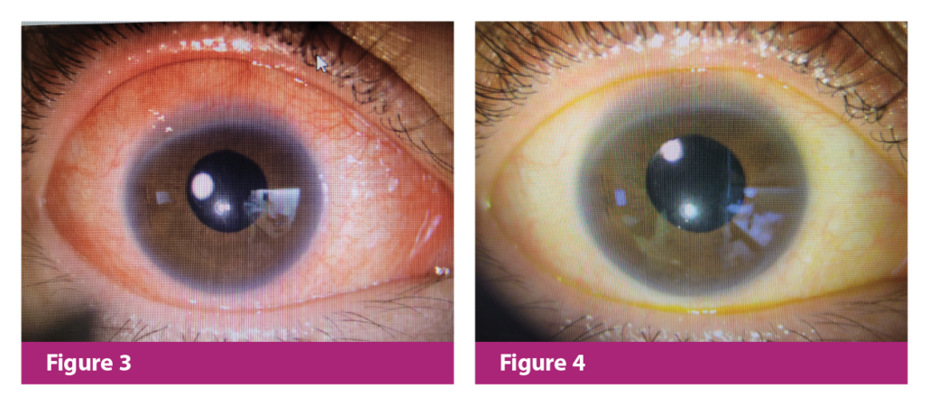
Figure 3. Right eye before treatment
Figure 4. Right eye 1 week after SLT and medication adjustment
Key Takeaways
Long-term administration of AGM eyedrops may lead to OSD due to prolonged exposure to preservatives and the active ingredients. The worsening of ocular surface will cause discomfort, and may contribute to poor treatment adherence, which in turn compromises the treatment outcome. Therefore, it is essential to address AGM-induced OSD properly and early to avoid the progression of glaucoma and patient’s negative impression on glaucoma eyedrops.
References
- Bielory B & Bielory L. Atopic dermatitis and keratoconjunctivitis. Immunol Allergy Clin North Am. 2010;30(3):323-36.
- Yang B, et al. Quantitative evaluation of lipid layer thickness and blinking in children with allergic conjunctivitis. Graefes Arch Clin Exp Ophthalmol. 2021;259(9):2795-805.
- Power WJ, et al. Long-term follow-up of patients with atopic keratoconjunctivitis. Ophthalmology. 1998;105(4):637-42.
- Kim SE, et al. Systemic interventions for severe atopic and vernal keratoconjunctivitis in children and young people up to the age of 16 years. Cochrane Database Syst Rev. 2020;10(10):CD013298.
- Ukponmwan CU. Vernal keratoconjunctivitis in Nigerians: 109 consecutive cases. Trop Doct. 2003;33(4):242-5.
- Hossain IT, et al. Pharmacotherapeutic management of atopic keratoconjunctivitis. Expert Opin Pharmacother. 2020;21(14):1761-9.
- Fineide F, et al. Topical glaucoma medications – Clinical implications for the ocular surface. The Ocul Surf. 2022;26:19-49.
- Andole S & Senthil S. Ocular Surface Disease and Anti-Glaucoma Medications: Various features, Diagnosis, and Management Guidelines. Semin Ophthalmol. 2023;38(2):158-66.
- Voicu L & Salim S. New strategies for the management of ocular surface disease in glaucoma patients. Curr Opin Ophthalmol. 2021;32(2):134-40.
- Fechtner RD, et al. Prevalence of ocular surface complaints in patients with glaucoma using topical intraocular pressure-lowering medications. Cornea. 2010;29(6):618-21.
- Kim JH, et al. Eyelid Changes Related to Meibomian Gland Dysfunction in Early Middle-Aged Patients Using Topical Glaucoma Medications. Cornea. 2018;37(4):421-5.
- Datta S, et al. The Eye Drop Preservative Benzalkonium Chloride Potently Induces Mitochondrial Dysfunction and Preferentially Affects LHON Mutant Cells. Invest Ophthalmol Vis Sci. 2017;58(4):2406-12.
- Zhang X, et al. Ocular Surface Disease and Glaucoma Medications: A Clinical Approach. Eye Contact Lens. 2019;45(1):11-8.

Editor
MediPaper Medical Communications Ltd
Unit 1104, 11/F Crawford house
70 Queen’s Road
Central, Hong Kong SAR
(852) 3008 1717 | medipr.org
FOTE-HK-230006
Disclaimer
This article is not medical advice. Patients should seek personal assessment by a licenced specialist. Physicians are recommended to read the full publication(s) as cited in the article before making medical decisions. This article does not supersede nor replace the published article(s).
© Copyright 2023 MediPaper Medical Communications Ltd. – HK Ocular Surface Newsletter Issue No. 2 Mar 2023
YOU MAY ALSO LIKE
© Copyright 2023 MediPaper Medical Communications Ltd. – HK Ocular Surface Newsletter Issue No. 2 Mar 2023


Leave a Reply
Want to join the discussion?Feel free to contribute!