![Medical News Asia | Powered by ['mediPr]](https://medi-paper.com/wp-content/uploads/2020/02/MediPR-MediPaper-Medical-Communications-Ltd-Healthcare-Writers-Medical-Writers-Medical-Writer-Agency-Hong-Kong-Medical-Writing-Services-Solutions-Quote-醫學寫作-香港-醫學作家香港-medical-PR-public-relations-1030x374.png)
HER2 Explorer
HER2 Unraveled—The Vital Distinction Between Overexpression and Gene Amplification in Cancer
Last updated: 11 December 2025 | In: HER2, HER2 Explorer
Significance of HER2 overexpression and HER2 amplification
Human epidermal growth factor receptor 2 (HER2) is a crucial biomarker in contemporary cancer management.11. Zhu K, et al. Biomark Res. 2024;12(1):16. Alterations in the HER2 protein or in the protein-coding gene, erythroblastic leukemia viral oncogene homolog 2 (ERBB2; hereafter referred to as HER2 in line with common practice), have significant clinical implications.11. Zhu K, et al. Biomark Res. 2024;12(1):16. Overexpression of the HER2 protein, gene amplification or activating mutations can overactivate or enhance downstream HER2 signaling pathways.2,3 2. Yoon J, Oh DY. Nat Rev Clin Oncol. 2024;21(9):675–700.
3. Chmielecki J, et al. Oncologist. 2015;20(1):7–12. Increased HER2 signaling drives key processes in tumorigenesis, including cellular transformation, survival, proliferation, and invasion.2,3 2. Yoon J, Oh DY. Nat Rev Clin Oncol. 2024;21(9):675–700.
3. Chmielecki J, et al. Oncologist. 2015;20(1):7–12.
Among these alterations, overexpression and gene amplification are closely related.11. Zhu K, et al. Biomark Res. 2024;12(1):16. Amplification refers to an increase in HER2 gene copy number, which often leads to overexpression, defined as an increased abundance of HER2 receptors on the cell surface (Figure 1).1,2,4-8 1. Zhu K, et al. Biomark Res. 2024;12(1):16.
2. Yoon J, Oh DY. Nat Rev Clin Oncol. 2024;21(9):675–700.
4. Connell CM, Doherty GJ. ESMO Open. 2017;2(5):e000279.
5. Hanna WM, et al. Mod Pathol. 2014;27(1):4–18.
6. Hechtman JF, et al. Cancer Cytopathol. 2019;127(7):428–31.
7. Neve RM, et al. Ann Oncol. 2001;12 Suppl 1:S9–13.
8. Yan M, et al. Cancer Metastasis Rev. 2015;34(1):157–64. However, gene amplification does not always correlate with protein overexpression, which can also occur through other mechanisms such as transcriptional regulation and post-transcriptional events.9,10 9. Nassar A, et al. Int J Clin Exp Pathol. 2014;7(9):6254–61.
10. Luoh SW, et al. Springerplus. 2013;2:386. Biological differences between HER2-overexpressing and HER2-amplified tumors remain poorly understood due to limited data.1111. Horimoto Y, et al. BMC Cancer. 2022;22(1):242.
In gastric and breast cancers, HER2 amplification is associated with poor prognosis and aggressive disease features, including shorter time to relapse and a higher propensity for brain metastases.12,1312. Pillai RN, et al. Cancer. 2017;123(21):4099–4105.
13. Iqbal N, Iqbal N. Mol Biol Int. 2014;2014:852748. An analysis of a phase III study suggests that the HER2-directed antibody trastuzumab may be less effective in HER2-amplified breast cancers compared with HER2-overexpressing tumors, likely due to the higher receptor abundance in the latter.11 11. Horimoto Y, et al. BMC Cancer. 2022;22(1):242.
An accurate assessment of HER2 status is important in the diagnostic evaluation of patients with solid tumors.1 1. Zhu K, et al. Biomark Res. 2024;12(1):16. For those whose tumors harbor HER2 alterations, multiple guideline-recommended HER2-targeted therapies are available, including monoclonal antibodies, antibody–drug conjugates (ADCs), and tyrosine kinase inhibitors (TKIs), with additional treatments in development.1 1. Zhu K, et al. Biomark Res. 2024;12(1):16. Therapy selection should be guided by the approved treatment indication, taking into account tumor type, specific alteration, and clinical context.14-1714. Bartley AN, et al. J Clin Oncol. 2017;35(4):446–64.
15. Wolff AC, et al. J Clin Oncol. 2018;36(20):2105–22.
16. Wolff AC, et al. J Clin Oncol. 2023;41(22):3867–72.
17. Daiichi Sankyo. ENHERTU® (fam-trastuzumab deruxtecan-nxki prescribing information. Available online. https://daiichisankyo.us/prescribing-information-portlet/getPIContent?productName=Enhertu&inline=true. Last accessed: 15 July 2025.
Currently, immunohistochemistry (IHC) and in situ hybridization (ISH) are the most commonly used methods to assess HER2 overexpression and gene amplification, respectively.1818. Troxell M, et al. Arch Pathol Lab Med. 2020;144(12):1525–34. In breast and gastric cancers, standardized IHC and ISH scoring criteria determine HER2 positivity, with IHC as the initial test and in situ hybridization (ISH) used for equivocal cases (IHC 2+).14-1614. Bartley AN, et al. J Clin Oncol. 2017;35(4):446–64.
15. Wolff AC, et al. J Clin Oncol. 2018;36(20):2105–22.
16. Wolff AC, et al. J Clin Oncol. 2023;41(22):3867–72. For other cancers, such as non-small cell lung cancer (NSCLC), molecular testing is recommended to detect activating HER2 mutations.1919. National Comprehensive Cancer Network. NCCN Clinical Practice Guidelines in Oncology – Non-Small Cell Lung Cancer Version 8.2025. Recently, HER2-targeted therapy has become available for patients with advanced or metastatic solid tumors meeting the IHC 3+ criteria, and, in these patients, confirming the presence of HER2 amplification is not necessary.1717. Daiichi Sankyo. ENHERTU® (fam-trastuzumab deruxtecan-nxki prescribing information. Available online. https://daiichisankyo.us/prescribing-information-portlet/getPIContent?productName=Enhertu&inline=true. Last accessed: 15 July 2025. Because the type of the HER2 alteration present in the tumor cells may impact a patient’s eligibility for targeted therapy,14-1714. Bartley AN, et al. J Clin Oncol. 2017;35(4):446–64.
15. Wolff AC, et al. J Clin Oncol. 2018;36(20):2105–22.
16. Wolff AC, et al. J Clin Oncol. 2023;41(22):3867–72.
17. Daiichi Sankyo. ENHERTU® (fam-trastuzumab deruxtecan-nxki prescribing information. Available online. https://daiichisankyo.us/prescribing-information-portlet/getPIContent?productName=Enhertu&inline=true. Last accessed: 15 July 2025.selecting the appropriate assessment method is essential to guide patients toward optimal therapeutic options.

Figure 1. Definition and testing methods of HER2 gene amplification and HER2 overexpression.
Prevalence of HER2 overexpression and HER2 amplification in solid tumors
The prevalence of HER2 overexpression and HER2 amplification varies widely across tumor types (Figure 2).22. Yoon J, Oh DY. Nat Rev Clin Oncol. 2024;21(9):675–700. Amplification of the HER2 gene and HER2 overexpression as defined by the IHC 3+ criteria are found in 20% and 10% of breast cancers, 40% and 15–37% of salivary gland cancers and 2% and 3% of lung cancers, respectively.22. Yoon J, Oh DY. Nat Rev Clin Oncol. 2024;21(9):675–700. This variability between amplification and overexpression in different solid tumors underscores the importance of tumor-specific testing strategies to guide appropriate HER2-directed therapy.
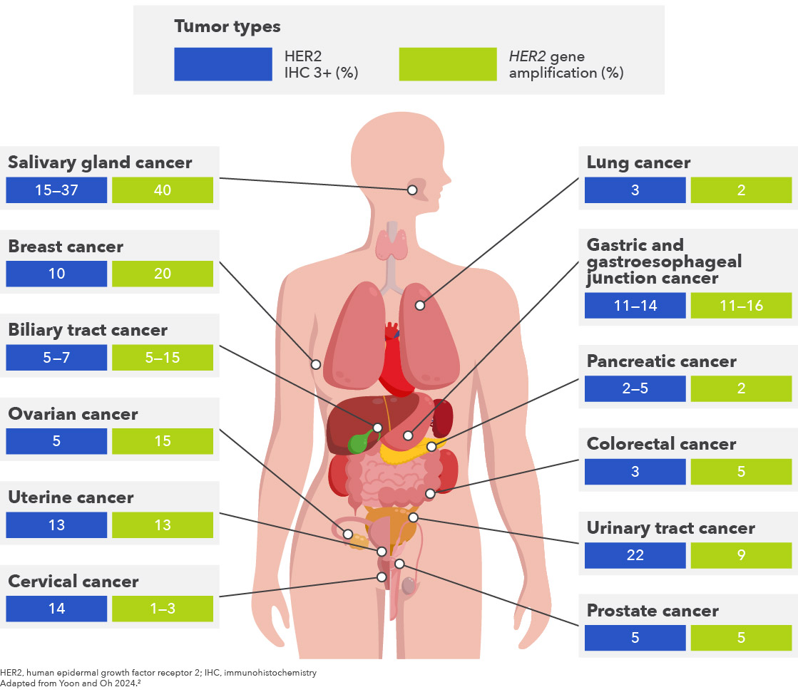
Figure 2: Prevalence of HER2 amplification and HER2 overexpression by tumor types.
Current standards and interpretation of HER2 testing guidelines for overexpression and amplification
Different diagnostic tests are required to identify different types of HER2 alterations, a topic covered in detail in article 5 of this series. IHC uses an anti-HER2 antibody as a probe to visualize HER2 protein abundance in a biopsy or surgical specimen and thereby detecting overexpression.55. Hanna WM, et al. Mod Pathol. 2014;27(1):4–18. Amplification of the HER2 gene is detected by ISH using a DNA probe that can detect increases in gene copy number.55. Hanna WM, et al. Mod Pathol. 2014;27(1):4–18. The DNA probes used in ISH may be coupled to a fluorescent (FISH), chromogenic (CISH) or silver (SISH) detection system.55. Hanna WM, et al. Mod Pathol. 2014;27(1):4–18. Single-probe ISH can measure HER2 copies per nucleus, and dual-probe ISH using an additional probe chromosome 17 centromere protein (CEP17) can determine the HER2:CEP17 ratio.55. Hanna WM, et al. Mod Pathol. 2014;27(1):4–18. The HER2:CEP17 ratio is considered to be a more accurate measure of amplification status as it is less affected by the mitotic index of the specimen, section thickness, and other potential sources of error.55. Hanna WM, et al. Mod Pathol. 2014;27(1):4–18. Recommendations for HER2 testing are available from the National Comprehensive Cancer Network (NCCN), the American Society of Clinical Oncology (ASCO)-College of American Pathologists (CAP), and other expert groups (Table 1).4,14,19,21-45 4. Connell CM, Doherty GJ. ESMO Open. 2017;2(5):e000279.
14. Bartley AN, et al. J Clin Oncol. 2017;35(4):446–64.
19. National Comprehensive Cancer Network. NCCN Clinical Practice Guidelines in Oncology – Non-Small Cell Lung Cancer Version 8.2025.
21. College of American Pathologists. Template for Reporting Results of Biomarker Testing of Specimens from Patients with Carcinoma of the Breast. Available online. https://documents.cap.org/documents/New-Cancer-Protocols-June-2025/Breast.Bmk_1.6.1.0.-REL_CAPCP.pdf. Last accessed: 27 November 2025.
22. College of American Pathologists. Template for Reporting Results of Biomarker Testing of Specimens From Patients With Carcinoma of Gynecologic Origin. Available online. https://documents.cap.org/protocols/Gynecologic.Bmk_1.3.0.0.REL_CAPCP.pdf. Last accessed: 27 November 2025.
23. College of American Pathologists. Template for Reporting Results of HER2 (ERBB2) Biomarker Testing of Specimens from Patients with Adenocarcinoma of the Stomach or Gastroesophageal Junction. Available online. https://documents.cap.org/protocols/cp-gastric-HER2biomarker17-1001.pdf. Last accessed: 27 November 2025.
24. College of American Pathologists. Template for Reporting Results of Biomarker Testing of Specimens From Patients With Carcinoma of the Colon and Rectum. Available online. https://documents.cap.org/protocols/ColoRectal.Bmk_1.3.0.0.REL_CAPCP.pdf. Last accessed: 27 November 2025.
25. College of American Pathologists. Template for Reporting Results of Biomarker Testing of Specimens From Patients With Non-Small Cell Carcinoma of the Lung. Available online. https://documents.cap.org/protocols/Lung.Bmk_2.2.0.0.REL_CAPCP.pdf. Last accessed: 27 November 2025.
26. College of American Pathologists. Head and Neck Biomarker Reporting Template. Available online. https://documents.cap.org/protocols/HN.Bmk_2.2.0.0.REL_CAPCP.pdf. Last accessed: 27 November 2025.
27. College of American Pathologists. Template for Reporting Results of Quantitative IHC Biomarker Testing of Specimens From Patients With Carcinoma. Available online. https://documents.cap.org/protocols/IHC.Bmk_1.2.0.0.REL_CAPCP.pdf Last accessed: 27 November 2025.
28. Fader AN, et al. J Clin Oncol. 2018;36(20):2044–51.
29. Goldsmith JD, et al. Arch Pathol Lab Med. 2024;148(6):e111–53.
30. National Comprehensive Cancer Network. NCCN Clinical Practice Guidelines in Oncology – Biliary Tract Cancers Version 2.2025.
31. National Comprehensive Cancer Network. NCCN Clinical Practice Guidelines in Oncology – Bladder Cancer Version 2.2025.
32. National Comprehensive Cancer Network. NCCN Clinical Practice Guidelines in Oncology – Ovarian Cancer Including Fallopian Tube Cancer and Primary Peritoneal Cancer Version 3.2025.
33. National Comprehensive Cancer Network. NCCN Clinical Practice Guidelines in Oncology – Cervical Cancer Version 2.2026.
34. National Comprehensive Cancer Network. NCCN Clinical Practice Guidelines in Oncology – Colon Cancer Version 5.2025.
35. National Comprehensive Cancer Network. NCCN Clinical Practice Guidelines in Oncology – Uterine Neoplasms Version 2.2026.
36. National Comprehensive Cancer Network. NCCN Clinical Practice Guidelines in Oncology – Gastric Cancer Version 3.2025.
37. National Comprehensive Cancer Network. NCCN Clinical Practice Guidelines in Oncology – Breast Cancer Version 5.2025.
38. National Comprehensive Cancer Network. NCCN Clinical Practice Guidelines in Oncology – Pancreatic Adenocarcinoma Version 2.2025.
39. National Comprehensive Cancer Network. NCCN Clinical Practice Guidelines in Oncology – Head and Neck Cancers Version 5.2025.
40. National Comprehensive Cancer Network. NCCN Clinical Practice Guidelines in Oncology – Prostate Cancer Version 3.2026.
41. National Comprehensive Cancer Network. NCCN Clinical Practice Guidelines in Oncology – Rectal Cancer Version 4.2025.
42. National Comprehensive Cancer Network. NCCN Clinical Practice Guidelines in Oncology – Esophageal and Esophagogastric Junction Cancers Version 4.2025.
43. Sartore-Bianchi A, et al. Lancet Oncol. 2016;17(6):738–46.
44. Zhang H, et al. Histopathology. 2024;85(1):3–19.
45. Wolff AC, et al. Arch Pathol Lab Med. 2023;147(9):993–1000
Specific and detailed scoring criteria for the interpretation of both ISH and IHC assays are well established for breast and gastric cancer.14,1514. Bartley AN, et al. J Clin Oncol. 2017;35(4):446–64.
15.Wolff AC, et al. J Clin Oncol. 2018;36(20):2105–22. For other solid tumors, a general HER2 IHC scoring template, which adopts the gastric HER2 IHC scoring, provided by the CAP is available to guide assessment.2727. College of American Pathologists. Template for Reporting Results of Quantitative IHC Biomarker Testing of Specimens From Patients With Carcinoma. Available online. https://documents.cap.org/protocols/IHC.Bmk_1.2.0.0.REL_CAPCP.pdf Last accessed 27 November 2025. Additionally, specific IHC scoring criteria have been developed and utilized as enrollment criteria in clinical trials.28,43 28. Fader AN, et al. J Clin Oncol. 2018;36(20):2044–51.
43. Sartore-Bianchi A, et al. Lancet Oncol. 2016;17(6):738–46. The appropriate testing modality varies by tumor type and may be impacted by tissue availability.

Table 1. Summary of HER2 testing recommendations in guidelines.4,14,19,21-45 4. Connell CM, Doherty GJ. ESMO Open. 2017;2(5):e000279.
14. Bartley AN, et al. J Clin Oncol. 2017;35(4):446–64.
19. National Comprehensive Cancer Network. NCCN Clinical Practice Guidelines in Oncology – Non-Small Cell Lung Cancer Version 8.2025.
21. College of American Pathologists. Template for Reporting Results of Biomarker Testing of Specimens from Patients with Carcinoma of the Breast. Available online. https://documents.cap.org/documents/New-Cancer-Protocols-June-2025/Breast.Bmk_1.6.1.0.-REL_CAPCP.pdf. Last accessed: 27 November 2025.
22. College of American Pathologists. Template for Reporting Results of Biomarker Testing of Specimens From Patients With Carcinoma of Gynecologic Origin. Available online. https://documents.cap.org/protocols/Gynecologic.Bmk_1.3.0.0.REL_CAPCP.pdf. Last accessed: 27 November 2025.
23. College of American Pathologists. Template for Reporting Results of HER2 (ERBB2) Biomarker Testing of Specimens from Patients with Adenocarcinoma of the Stomach or Gastroesophageal Junction. Available online. https://documents.cap.org/protocols/cp-gastric-HER2biomarker17-1001.pdf. Last accessed: 27 November 2025.
24. College of American Pathologists. Template for Reporting Results of Biomarker Testing of Specimens From Patients With Carcinoma of the Colon and Rectum. Available online. https://documents.cap.org/protocols/ColoRectal.Bmk_1.3.0.0.REL_CAPCP.pdf. Last accessed: 27 November 2025.
25. College of American Pathologists. Template for Reporting Results of Biomarker Testing of Specimens From Patients With Non-Small Cell Carcinoma of the Lung. Available online. https://documents.cap.org/protocols/Lung.Bmk_2.2.0.0.REL_CAPCP.pdf. Last accessed: 27 November 2025.
26. College of American Pathologists. Head and Neck Biomarker Reporting Template. Available online. https://documents.cap.org/protocols/HN.Bmk_2.2.0.0.REL_CAPCP.pdf. Last accessed: 27 November 2025.
27. College of American Pathologists. Template for Reporting Results of Quantitative IHC Biomarker Testing of Specimens From Patients With Carcinoma. Available online. https://documents.cap.org/protocols/IHC.Bmk_1.2.0.0.REL_CAPCP.pdf Last accessed: 27 November 2025.
28. Fader AN, et al. J Clin Oncol. 2018;36(20):2044–51.
29. Goldsmith JD, et al. Arch Pathol Lab Med. 2024;148(6):e111–53.
30. National Comprehensive Cancer Network. NCCN Clinical Practice Guidelines in Oncology – Biliary Tract Cancers Version 2.2025.
31. National Comprehensive Cancer Network. NCCN Clinical Practice Guidelines in Oncology – Bladder Cancer Version 2.2025.
32. National Comprehensive Cancer Network. NCCN Clinical Practice Guidelines in Oncology – Ovarian Cancer Including Fallopian Tube Cancer and Primary Peritoneal Cancer Version 3.2025.
33. National Comprehensive Cancer Network. NCCN Clinical Practice Guidelines in Oncology – Cervical Cancer Version 2.2026.
34. National Comprehensive Cancer Network. NCCN Clinical Practice Guidelines in Oncology – Colon Cancer Version 5.2025.
35. National Comprehensive Cancer Network. NCCN Clinical Practice Guidelines in Oncology – Uterine Neoplasms Version 2.2026.
36. National Comprehensive Cancer Network. NCCN Clinical Practice Guidelines in Oncology – Gastric Cancer Version 3.2025.
37. National Comprehensive Cancer Network. NCCN Clinical Practice Guidelines in Oncology – Breast Cancer Version 5.2025.
38. National Comprehensive Cancer Network. NCCN Clinical Practice Guidelines in Oncology – Pancreatic Adenocarcinoma Version 2.2025.
39. National Comprehensive Cancer Network. NCCN Clinical Practice Guidelines in Oncology – Head and Neck Cancers Version 5.2025.
40. National Comprehensive Cancer Network. NCCN Clinical Practice Guidelines in Oncology – Prostate Cancer Version 3.2026.
41. National Comprehensive Cancer Network. NCCN Clinical Practice Guidelines in Oncology – Rectal Cancer Version 4.2025.
42. National Comprehensive Cancer Network. NCCN Clinical Practice Guidelines in Oncology – Esophageal and Esophagogastric Junction Cancers Version 4.2025.
43. Sartore-Bianchi A, et al. Lancet Oncol. 2016;17(6):738–46.
44. Zhang H, et al. Histopathology. 2024;85(1):3–19.
45. Wolff AC, et al. Arch Pathol Lab Med. 2023;147(9):993–1000
HER2 ISH scoring criteria in breast and gastric cancers
For patients with breast or gastric cancer, HER2 status needs to be accurately evaluated to identify patients who may be eligible for targeted therapies. This may require the use of both IHC and ISH. Detailed IHC scoring guidelines for breast and gastric cancers are available from ASCO-CAP,14,1514. Bartley AN, et al. J Clin Oncol. 2017;35(4):446–64.
15. Wolff AC, et al. J Clin Oncol. 2018;36(20):2105–22. and these have been reviewed in detail previously. These guidelines also make recommendations for ISH scoring for these two tumor types, with dual-probe ISH as the preferred approach for breast cancer and the only approach recommended for gastric cancer (Figure 3).14,15,17,21,36,37,4514. Bartley AN, et al. J Clin Oncol. 2017;35(4):446–64.
15. Wolff AC, et al. J Clin Oncol. 2018;36(20):2105–22.
17. Daiichi Sankyo. ENHERTU® (fam-trastuzumab deruxtecan-nxki prescribing information. Available online. https://daiichisankyo.us/prescribing-information-portlet/getPIContent?productName=Enhertu&inline=true. Last accessed: 15 July 2025.
21. College of American Pathologists. Template for Reporting Results of Biomarker Testing of Specimens from Patients with Carcinoma of the Breast. Available online. https://documents.cap.org/documents/New-Cancer-Protocols-June-2025/Breast.Bmk_1.6.1.0.-REL_CAPCP.pdf. Last accessed: 27 November 2025.
36. National Comprehensive Cancer Network. NCCN Clinical Practice Guidelines in Oncology – Gastric Cancer Version 3.2025.
37. National Comprehensive Cancer Network. NCCN Clinical Practice Guidelines in Oncology – Breast Cancer Version 5.2025.
45. Wolff AC, et al. Arch Pathol Lab Med. 2023;147(9):993–1000. For patients with breast cancer, ISH scores are categorized into five groups based on HER2:CEP17 ratio and average number of signals per cell.1515. Wolff AC, et al. J Clin Oncol. 2018;36(20):2105–22. Groups 1 and 5 (HER2 positive and negative, respectively) account for ~95% of tumors tested.1515. Wolff AC, et al. J Clin Oncol. 2018;36(20):2105–22. Specimens in Groups 2–4 need additional data from IHC testing to confirm their HER2 status.1515. Wolff AC, et al. J Clin Oncol. 2018;36(20):2105–22. For those with gastric cancer, single probe ISH is not recommended.3636. National Comprehensive Cancer Network. NCCN Clinical Practice Guidelines in Oncology – Gastric Cancer Version 3.2025. HER2 positive is defined as HER2:CEP17 ratio ≥2.0 or an average HER2 copy number of ≥6 signals per cell.3636. National Comprehensive Cancer Network. NCCN Clinical Practice Guidelines in Oncology – Gastric Cancer Version 3.2025. In summary, assessment of HER2 status with both ISH and IHC is vital to ensure that all breast and gastric cancer patients eligible for HER2-directed therapies are identified.

Figure 3. Testing and interpretation of HER2 ISH in breast and gastric cancer
Concordance between IHC and ISH evaluation of HER2
Despite the mechanistic relationship between gene amplification and protein overexpression, the concordance between IHC and ISH evaluation of HER2 is not absolute. The reported concordance between IHC 3+ and ISH testing is 91% for both breast and gastric cancers.46,4746. Pyo JS, et al. Int J Biol Markers. 2016;31(1):e1–10.
47. Bahreini F, et al. Breast Cancer. 2015;22(6):615–25. Polysomy 17 is the likely cause of this inconsistency.99. Nassar A, et al. Int J Clin Exp Pathol. 2014;7(9):6254–61. Conversely, some breast cancers test positive for HER2 gene amplification but do not over-express HER2; these patients are less likely to benefit from HER2-directed therapies.99. Nassar A, et al. Int J Clin Exp Pathol. 2014;7(9):6254–61.
HER2 Amplification Assessment via NGS — Opportunities and Limitations
Although ISH is the guideline-recommended standard for detecting HER2 gene amplifications in breast, gastric, and other cancers,14,1514. Bartley AN, et al. J Clin Oncol. 2017;35(4):446–64.
15. Wolff AC, et al. J Clin Oncol. 2018;36(20):2105–22. the use of next-generation sequencing (NGS) to detect mutations and amplifications is recommended in guidelines for some tumor types (e.g., NSCLC and colorectal cancer),19,34,4119. National Comprehensive Cancer Network. NCCN Clinical Practice Guidelines in Oncology – Non-Small Cell Lung Cancer Version 8.2025.
34. National Comprehensive Cancer Network. NCCN Clinical Practice Guidelines in Oncology – Colon Cancer Version 5.2025.
41. National Comprehensive Cancer Network. NCCN Clinical Practice Guidelines in Oncology – Rectal Cancer Version 4.2025. and there is increasing interest in this approach. NGS allows the assessment of large numbers of genes and can make more efficient use of limited biopsy specimens or permit the use of liquid biopsies. However, challenges exist in the concordance between NGS estimates of gene amplification and ISH. A study comparing tissue-based NGS and dual-probe ISH for measuring HER2 amplifications for breast cancer determined NGS cutoff values (copy number >4.09 and <2.91) for HER2 amplification status that had 95% sensitivity and 99% specificity with reference to ISH.4848. Niu D, et al. Pathol Oncol Res. 2020;26(4):2577–85. However, the same cutoff had lower sensitivity (65%) with gastric cancer specimens, possibly due to tumor heterogeneity.4848. Niu D, et al. Pathol Oncol Res. 2020;26(4):2577–85. Similarly, a study by Shah et al. that included multiple tumor types found varying concordance between copy number gain by NGS and standard IHC/ISH definitions of positivity by tumor and specimen type.4949. Shah AN, et al. Oncotarget. 2022;13:273–80. Tissue NGS and circulating tumor DNA (ctDNA) NGS positive predictive value (PPV) for HER2 positivity by IHC/ISH was highest for breast cancer, 97% and 93% respectively, but PPV was lower for non-breast cancer tumors (76% tissue, 44% ctDNA).4949. Shah AN, et al. Oncotarget. 2022;13:273–80. Considerable discordance may exist between NGS and tissue IHC/ISH. For example, in two studies of biliary tract cancer, both using the same ctDNA NGS assay, positive percent agreement between ctDNA NGS and tissue IHC/ISH varied between 36.4% and 75.9%.50,5150. Nakamura Y, et al. J Clin Oncol. 2023;41(36):5569–78.
51. Ohba A, et al. J Clin Oncol. 2023;41(16_suppl):4097. Further research should be conducted to determine appropriate diagnostic cutoff values for different NGS panels in specific tumor types.
Precision in HER2 testing allows targeted treatment
Due to the discordance of HER2 status assessed by IHC, ISH, or NGS,4949. Shah AN, et al. Oncotarget. 2022;13:273–80. HER2 gene amplification and HER2 overexpression should not be considered as clinically equivalent.99. Nassar A, et al. Int J Clin Exp Pathol. 2014;7(9):6254–61. NGS has the potential to serve as a tumor-agnostic approach for assessing HER2 amplification, but currently, concordance with established methods is highly variable.48,4948. Niu D, et al. Pathol Oncol Res. 2020;26(4):2577–85.
49. Shah AN, et al. Oncotarget. 2022;13:273–80. HER2 overexpression assessed by IHC remains the only HER2 alteration with a tumor-agnostic indication for HER2-targeted therapy.1717. Daiichi Sankyo. ENHERTU® (fam-trastuzumab deruxtecan-nxki prescribing information. Available online. https://daiichisankyo.us/prescribing-information-portlet/getPIContent?productName=Enhertu&inline=true. Last accessed: 15 July 2025. At present, HER2 testing strategies should be aligned with current guidelines, treatment indication and individualized clinical objectives.
References
- Zhu K, et al. Biomark Res. 2024;12(1):16.
- Yoon J, Oh DY. Nat Rev Clin Oncol. 2024;21(9):675–700.
- Chmielecki J, et al. Oncologist. 2015;20(1):7–12.
- Connell CM, Doherty GJ. ESMO Open. 2017;2(5):e000279.
- Hanna WM, et al. Mod Pathol. 2014;27(1):4–18.
- Hechtman JF, et al. Cancer Cytopathol. 2019;127(7):428–31.
- Neve RM, et al. Ann Oncol. 2001;12 Suppl 1:S9–13.
- Yan M, et al. Cancer Metastasis Rev. 2015;34(1):157–64.
- Nassar A, et al. Int J Clin Exp Pathol. 2014;7(9):6254–61.
- Luoh SW, et al. Springerplus. 2013;2:386.
- Horimoto Y, et al. BMC Cancer. 2022;22(1):242.
- Pillai RN, et al. Cancer. 2017;123(21):4099–4105.
- Iqbal N, Iqbal N. Mol Biol Int. 2014;2014:852748.
- Bartley AN, et al. J Clin Oncol. 2017;35(4):446–64.
- Wolff AC, et al. J Clin Oncol. 2018;36(20):2105–22.
- Wolff AC, et al. J Clin Oncol. 2023;41(22):3867–72.
- Daiichi Sankyo. ENHERTU® (fam-trastuzumab deruxtecan-nxki prescribing information. Available online. https://daiichisankyo.us/prescribing-information-portlet/getPIContent?productName=Enhertu&inline=true. Last accessed: 15 July 2025.
- Troxell M, et al. Arch Pathol Lab Med. 2020;144(12):1525–34.
- National Comprehensive Cancer Network. NCCN Clinical Practice Guidelines in Oncology – Non-Small Cell Lung Cancer Version 8.2025.
- Liu Q, et al. Oncogene. 2018;37(5):687–94.
- College of American Pathologists. Template for Reporting Results of Biomarker Testing of Specimens from Patients with Carcinoma of the Breast. Available online. https://documents.cap.org/documents/New-Cancer-Protocols-June-2025/Breast.Bmk_1.6.1.0.-REL_CAPCP.pdf. Last accessed: 27 November 2025.
- College of American Pathologists. Template for Reporting Results of Biomarker Testing of Specimens From Patients With Carcinoma of Gynecologic Origin. Available online. https://documents.cap.org/protocols/Gynecologic.Bmk_1.3.0.0.REL_CAPCP.pdf. Last accessed: 27 November 2025.
- College of American Pathologists. Template for Reporting Results of HER2 (ERBB2) Biomarker Testing of Specimens from Patients with Adenocarcinoma of the Stomach or Gastroesophageal Junction. Available online. https://documents.cap.org/protocols/cp-gastric-HER2biomarker17-1001.pdf. Last accessed: 27 November 2025.
- College of American Pathologists. Template for Reporting Results of Biomarker Testing of Specimens From Patients With Carcinoma of the Colon and Rectum. Available online. https://documents.cap.org/protocols/ColoRectal.Bmk_1.3.0.0.REL_CAPCP.pdf. Last accessed: 27 November 2025.
- College of American Pathologists. Template for Reporting Results of Biomarker Testing of Specimens From Patients With Non-Small Cell Carcinoma of the Lung. Available online. https://documents.cap.org/protocols/Lung.Bmk_2.2.0.0.REL_CAPCP.pdf. Last accessed: 27 November 2025.
- College of American Pathologists. Head and Neck Biomarker Reporting Template. Available online. https://documents.cap.org/protocols/HN.Bmk_2.2.0.0.REL_CAPCP.pdf. Last accessed: 27 November 2025.
- College of American Pathologists. Template for Reporting Results of Quantitative IHC Biomarker Testing of Specimens From Patients With Carcinoma. Available online. https://documents.cap.org/protocols/IHC.Bmk_1.2.0.0.REL_CAPCP.pdf. Last accessed: 27 November 2025.
- Fader AN, et al. J Clin Oncol. 2018;36(20):2044–51.
- Goldsmith JD, et al. Arch Pathol Lab Med. 2024;148(6):e111–53.
- National Comprehensive Cancer Network. NCCN Clinical Practice Guidelines in Oncology – Biliary Tract Cancers Version 2.2025.
- National Comprehensive Cancer Network. NCCN Clinical Practice Guidelines in Oncology – Bladder Cancer Version 2.2025.
- National Comprehensive Cancer Network. NCCN Clinical Practice Guidelines in Oncology – Ovarian Cancer Including Fallopian Tube Cancer and Primary Peritoneal Cancer Version 3.2025.
- National Comprehensive Cancer Network. NCCN Clinical Practice Guidelines in Oncology – Cervical Cancer Version 2.2026.
- National Comprehensive Cancer Network. NCCN Clinical Practice Guidelines in Oncology – Colon Cancer Version 5.2025.
- National Comprehensive Cancer Network. NCCN Clinical Practice Guidelines in Oncology – Uterine Neoplasms Version 2.2026.
- National Comprehensive Cancer Network. NCCN Clinical Practice Guidelines in Oncology – Gastric Cancer Version 3.2025.
- National Comprehensive Cancer Network. NCCN Clinical Practice Guidelines in Oncology – Breast Cancer Version 5.2025.
- National Comprehensive Cancer Network. NCCN Clinical Practice Guidelines in Oncology – Pancreatic Adenocarcinoma Version 2.2025.
- National Comprehensive Cancer Network. NCCN Clinical Practice Guidelines in Oncology – Head and Neck Cancers Version 5.2025.
- National Comprehensive Cancer Network. NCCN Clinical Practice Guidelines in Oncology – Prostate Cancer Version 3.2026.
- National Comprehensive Cancer Network. NCCN Clinical Practice Guidelines in Oncology – Rectal Cancer Version 4.2025.
- National Comprehensive Cancer Network. NCCN Clinical Practice Guidelines in Oncology – Esophageal and Esophagogastric Junction Cancers Version 4.2025.
- Sartore-Bianchi A, et al. Lancet Oncol. 2016;17(6):738–46.
- Zhang H, et al. Histopathology. 2024;85(1):3–19.
- Wolff AC, et al. Arch Pathol Lab Med. 2023;147(9):993–1000.
- Pyo JS, et al. Int J Biol Markers. 2016;31(1):e1–10.
- Bahreini F, et al. Breast Cancer. 2015;22(6):615–25.
- Niu D, et al. Pathol Oncol Res. 2020;26(4):2577–85.
- Shah AN, et al. Oncotarget. 2022;13:273–80.
- Nakamura Y, et al. J Clin Oncol. 2023;41(36):5569–78.
- Ohba A, et al. J Clin Oncol. 2023;41(16_suppl):4097.
The article is sponsored by Daiichi Sankyo and AstraZeneca.
Disclaimer
This article is not medical advice. Patients should seek personal assessment by a licenced specialist. Physicians are recommended to read the full publication(s) as cited in the article before making medical decisions. This article does not supersede nor replace the published article(s).
© Copyright 2025 MediPaper Medical Communications Ltd. – HER2 Unraveled—The Vital Distinction Between Overexpression and Gene Amplification in Cancer

