![Medical News Asia | Powered by ['mediPr]](https://medi-paper.com/wp-content/uploads/2020/02/MediPR-MediPaper-Medical-Communications-Ltd-Healthcare-Writers-Medical-Writers-Medical-Writer-Agency-Hong-Kong-Medical-Writing-Services-Solutions-Quote-醫學寫作-香港-醫學作家香港-medical-PR-public-relations-1030x374.png)

OPHTHALMOLOGY
HK Ocular Surface Newsletter Issue No. 1 Nov 2022
Last updated: 18 November 2022 | In: Ophthalmology, Therapeutic Areas
Article Keywords
dry eye disease, meibomian gland dysfunction, ophthalmology, topical cyclosporine, vernal keratoconjunctivitis

Dr. ALEX L.K. NG
Specialist in Ophthalmology
The Hong Kong Ophthalmic Associates
When and How to Start Cyclosporine in Dry Eye Disease
The pathophysiology of dry eye disease (DED) is attributed to a cascade of inflammatory events that results in a vicious circle of immune responses.11. Jones L, et al. Ocul Surf. 2017;15(3):575-628. When patients are exposed to external factors or certain ocular stress, tear osmolarity could increase, leading to ocular surface inflammation, which plays a key role in DED and causes further damage to the corneal epithelial cells.1-31. Jones L, et al. Ocul Surf. 2017;15(3):575-628.
2. Potvin R, et al. Clin Ophthalmol. 2015;9:2039-47.
3. Soifer M, et al. Front Med (Lausanne). 2022;9:949202.
The Tear Film and Ocular Surface Society (TFOS) International Dry Eye Workshop II (DEWS II) report recommends treating inflammation at the relatively early stage. A stepwise approach is proposed depending on the patient’s severity of signs and symptoms. Many ophthalmologists had an impression that topical cyclosporine (CsA) is only initiated at a very late or severe stage. However, if we look at the DEWS II recommendations of staged management and the treatment recommendations for DED, topical CsA is actually recommended at step 2, alongside with other common treatment options such as topical corticosteroids.11. Jones L, et al. Ocul Surf. 2017;15(3):575-628.
“As most patients are concerned about the cost of drug and the treatment duration of 6 to 12 months, patient compliance could be a challenge in the prescription process. This creates a need for chair time for comprehensive patient education,” Dr. Ng explained. “This includes clarifying the role of inflammation in DED and the safety of topical CsA in addressing inflammation. It is also important to note that the onset of CsA action is not immediate. Patient should also be warned about the mild irritation or transient eye pain after instillation of CsA eyedrops, which is common, but mostly tolerable and patients should be reassured. However, the good side is topical CsA can be prescribed for long term to sustain the control of symptoms, unlike topical corticosteroids. The once-daily dosing of topical 0.1% CsA also allows for better patient compliance.4,54. Baudouin C, et al. Eur J Ophthalmol. 2017;27(6):678-855. Ikervis® 1mg/mL Hong Kong Package Insert. 2018.”
Case Sharing
Case 1: Aqueous Deficient Dry Eye with Significant Staining
Dr. Ng shared a case presenting with typical dry eye associated severe keratitis, that he would prefer prescribing topical 0.1% CsA right away. It involved a 50-year-old female patient with known rheumatoid arthritis (RA) and secondary Sjögren’s syndrome (SS), with anti-Sjögren’s-syndrome-related antigen A (anti-SSA) autoantibodies positive, presenting with dryness (++), and blurred vision. It is a typical example of aqueous deficient dry eye, additionally presenting with significant staining (+++) on the ocular surface (Figures 1 & 2).
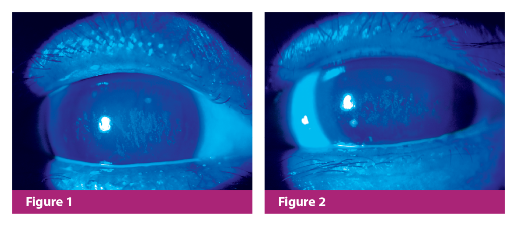
Figure 1. Corneal staining of the right eye before topical CsA treatment
Figure 2. Corneal staining of the left eye before topic CsA treatment
The patient was started with topical 0.1% CsA right away, together with fuorometholone ophthalmic suspension (FML) tapered over 6-8 weeks, artificial tears, and collagen plugs. At the 6-month follow-up, the patient was nearly symptom-free despite mild residual staining (Figures 3 & 4). Due to her medical history of RA and SS, long-term use of topical 0.1% CsA was recommended.
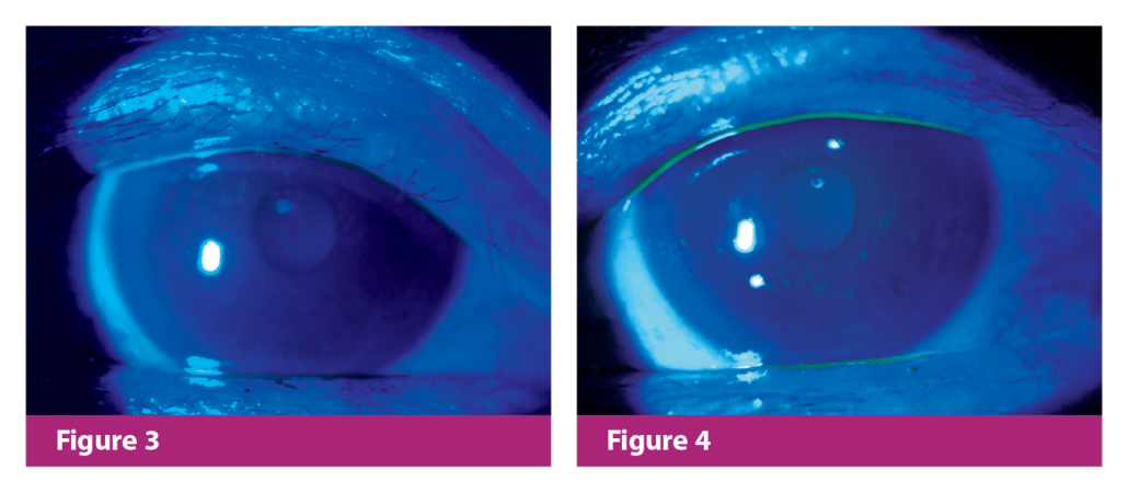
Figure 3. Corneal staining of the right eye after topical CsA treatment
Figure 4. Corneal staining of the left eye after topical CsA treatment
Dr. Ng mentioned that although the classical indication for topical 0.1% CsA was severe keratitis, there was a consensus among ocular surface disease experts to initiate CsA at an earlier stage. Topical CsA can be started in patients with milder grades of corneal fuorescein staining (CFS), depending on other factors such as how symptomatic the patient is, and whether patients have received any prior DED treatments.
Case 2: Evaporative Dry Eye induced by MGD without Staining
The second case is a 24-year-old female patient with a 1-year history of recurrent inflammation with redness, pain, and dryness. She was previously diagnosed with DED due to meibomian gland dysfunction (MGD), treated with lubricants, warm compresses, and tea tree oil for DED and MGD, as well as topical corticosteroids in each inflammation episode.
The patient consulted Dr. Ng after a recurrence of inflammation in her right eye, with redness and mild pain (Figure 5). Clinical examination confirmed a significant MGD with a short tear breakup time (TBUT), with injection++ in the right eye. There was no corneal staining. It was also revealed from history taking that the MGD onset coincided with her retinoic acid treatment for acne.

Figure 5. Meibomian gland dysfunction and inflammation of the eye
Considering the frequent occurrence of inflammatory episodes requiring steroids, Dr. Ng initiated topical 0.1% CsA on top of topical corticosteroids for this episode, aiming to reduce the occurrence of future inflammatory episodes and thus steroid use. Her MGD condition improved after LipiFlow treatment and the termination of retinoic acid treatment, with only one mild redness episode which required several days of treatment with FML. The patient remained free of DED and ocular surface inflammation for months when she was only on topical 0.1% CsA treatment and warm compresses. This example highlights another spectrum of DED patients with recurrent episodes of inflammation, where topical CsA helps reduce steroid-dependence and restore a stable ocular surface.
Key Takeaway Messages
Dr. Ng highlighted the importance of addressing ocular surface inflammation at an earlier stage when managing DED. To increase patients’ treatment compliance, physicians should explain the rationale for topical CsA treatment and manage patient expectations, such as potential discomfort during instillation of eyedrops. Nevertheless, apart from treating inflammation, other contributing factors of DED should also be addressed.

Dr. DOUGLAS K.T. LAM
Specialist in Ophthalmology
The Hong Kong Ophthalmic Associates
Assessing and Treating Vernal Keratoconjunctivitis with Cyclosporine
Vernal keratoconjunctivitis (VKC) is an ocular inflammation commonly diagnosed in children aged 5 to 10. The most common ocular complications include redness, itching, increased mucus discharge, and blurred vision. It may occur seasonally due to pollen, or perennially from year-round exposure to allergens such as dust mites, which often involve acute exacerbation and recurrence.66. Mehta JS, et al. Front Med (Lausanne). 2022;9:882240.
The lack of well-defined, standardized guidelines on VKC management in the past resulted in variability in VKC diagnosis and treatment across Asia and among physicians. In view of this, a panel of experts from the Management of Vernal Keratoconjunctivitis in Asia (MOVIA) Expert Working Group, including Dr. Douglas Lam and Dr. Arthur Cheng from Hong Kong, developed a set of Asian VKC consensus of best-practice recommendations for the assessment, diagnosis, and management of VKC in 2022.66. Mehta JS, et al. Front Med (Lausanne). 2022;9:882240.
The consensus has clarified the diagnostic criteria based on the most common reference in Asia, the Bonini’s scale, and proposed stepwise VKC management based on the disease severity. Based on this scale, the severity of VKC is classified into quiescent, mild, moderate, severe, very severe, or evolution, according to increasing disturbance by symptoms, and increasing corneal involvement and complications, on top of the presence of giant papillae.66. Mehta JS, et al. Front Med (Lausanne). 2022;9:882240. Dr. Lam, as in the consensus, highlighted the significance of slit-lamp examination and eversion of eyelids during assessment, to better evaluate if the patient suffers from tarsal form, limbal form, or mixed form of VKC.
Stepwise approach management will include addressing triggers to the disease, e.g., environment and allergens, maintaining ocular health through frequent hand, face, and hair washing, as well as using ocular lubricants and cold compresses.
Regarding the pharmacological options, below are the highlights from the consensus:66. Mehta JS, et al. Front Med (Lausanne). 2022;9:882240.
- When using conventional topical anti-allergic drugs, dual-acting agents should be considered ahead of monotherapy with anti-histamines or mast cell stabilizers alone
- Topical 0.1% CsA should be considered for patients with moderate-to-severe or persistent VKC, with proper usage instructions to minimize stinging on instillation
- Topical corticosteroids are effective for management of acute exacerbation. They are preferentially used in more severe cases, including those with corneal involvement, or cases where only conjunctival involvement was observed, however with a loss of control or persistence of symptoms with immunomodulators (such as topical CsA)
- Tacrolimus should be reserved for patients with severe VKC that is refractory to topical CsA, with the note of possible off-label usage
- Vasoconstrictors and non-steroidal anti-inflammatory drugs (NSAIDS) are not recommended, with the respective reason of their adverse events and lacking the target of the specific inflammatory mechanism associated with VKC
Dr. Lam shared his routine of practice, with the first step of determining the disease severity. He differentiates mild VKC from moderate-to-severe VKC based on the presence of corneal or conjunctival involvement, like papillae or Horner-Trantas dots. Presentation of corneal or conjunctival involvement indicates moderate-to-severe VKC. While cases with the absence of the aforementioned conditions will be considered mild. Anti-histamines eye drops may be given to mild cases to relieve signs and symptoms. Topical 0.1% CsA may be prescribed for moderate cases with conjunctival involvement like papillae or limbal involvement. For severe or progressed cases with corneal complication like cornea ulcer, short-course corticosteroid treatment may be added to the topical 0.1% CsA treatment.6,76. Mehta JS, et al. Front Med (Lausanne). 2022;9:882240.7. Leonardi A. Ophthalmol Ther. 2013;2(2):73-88.
Dr. Lam further mentioned topical 0.1% CsA could achieve sustained control of signs and symptoms, and reduce the use of topical corticosteroids and its related side effects, such as raised intraocular pressure that may lead to glaucoma or cataract formation. “Therefore, the prescription of topical 0.1% CsA should be encouraged as a first-line treatment instead of topical corticosteroids,” Dr. Lam said.
Case Sharing
A 10-year-old boy with 2-year history of recurrent VKC and was pretreated only with topical corticosteroids was referred to Dr. Lam. Dr. Lam graded this case as a severe VKC in tarsal form, with corneal ulcer, bilateral redness, and discharge (Figures 6 & 7), which required immediate treatment to avoid sight-threatening complications. He was prescribed topical 0.1% CsA and a tapered 2 week preservative-free topical corticosteroid treatment simultaneously. A remarkable improvement was seen after 1.5 months of treatment. The papillae were minimised, and redness in the eyes was resolved (Figures 8). Dr. Lam continued to prescribe topical 0.1% CsA for long-term maintenance for the next 2 to 3 years, while titrated down gradually from QID to QD as the condition improved (Figure 9). With the effective and sustained control being achieved with topical 0.1% CsA, Dr. Lam would like to encourage peers to consider prescribing the drug as the first-line treatment for VKC.

Figure 6. Right upper eyelid before treatment
Figure 7. Right eye before treatment
Figure 8. Right upper eyelid, after 1.5 months of topical 0.1% CsA and tapering topical corticosteroid treatment
Figure 9. Right eye, after 2 years of topical 0.1% CsA maintenance treatment
Tips from Dr. Lam
As VKC has a high recurrence rate, topical 0.1% CsA can be a safe and effective option for long-term use.88. Leonardi A, et al. Ophthalmology. 2019;126(5):671-81. Quality of life is also an important consideration as VKC impacts daily activities, such as children’s social interactions and attendance at school.66. Mehta JS, et al. Front Med (Lausanne). 2022;9:882240. To maintain treatment compliance in school-going patients, a doctor’s note may be provided for the school nurse (or caregivers) to help administer the eye drops in school.
It is also worth noting that patient expectation management is essential, such as notifying patients’ caregivers about stinging sensations from CsA.
If topical corticosteroids are prescribed, doctors should closely monitor for ocular complications and taper down as soon as possible to minimize the complications.
References
- Jones L, et al. Ocul Surf. 2017;15(3):575-628.
- Potvin R, et al. Clin Ophthalmol. 2015;9:2039-47.
- Soifer M, et al. Front Med (Lausanne). 2022;9:949202.
- Baudouin C, et al. Eur J Ophthalmol. 2017;27(6):678-85.
- Ikervis® 1mg/mL Hong Kong Package Insert. 2018.
- Mehta JS, et al. Front Med (Lausanne). 2022;9:882240.
- Leonardi A. Ophthalmol Ther. 2013;2(2):73-88.
- Leonardi A, et al. Ophthalmology. 2019;126(5):671-81.

Editor
MediPaper Medical Communications Ltd
Unit 1104, 11/F Crawford house
70 Queen’s Road
Central, Hong Kong SAR
(852) 3008 1717 | medipr.org
FOTE-HK-220003
Disclaimer
This article is not medical advice. Patients should seek personal assessment by a licenced specialist. Physicians are recommended to read the full publication(s) as cited in the article before making medical decisions. This article does not supersede nor replace the published article(s).
© Copyright 2022 MediPaper Medical Communications Ltd. – HK Ocular Surface Newsletter Issue No. 1 Nov 2022
YOU MAY ALSO LIKE
© Copyright 2022 MediPaper Medical Communications Ltd. – HK Ocular Surface Newsletter Issue No. 1 Nov 2022


Leave a Reply
Want to join the discussion?Feel free to contribute!